Showing posts with label skeleton. Show all posts

Saturday, June 4, 2016

Current projects


Been a while since I posted anything here.
Still busy with our upcoming website, but in the meantime we have been working on a couple of really interesting projects, which are almost completed now, but I'll just post some early pictures for now.

We have been asked to clean a full shark and skate skeletons for a french natural history museum. This is a really exciting opportunity, and they look fantastic so far. The species are a school shark (Galeorhinus galeus) and undulate ray (Raja undulata). Both are male specimens caught off the coast of Brittany in France. The plan is to have them displayed next to their taxidermied versions, which were recently completed by Bernard Bourles, an excellent ichthyotaxidermist (his website can be found here: http://www.poissons.net/).

Galeorhinus galeus. This specimen is a male around 1.20 m long.

Dissection of the gills, this part needs to be done very carefully as not to damage the delicate branchial rays and extrabranchials, which are visible on top of the gills here.

Raja undulata, male specimen as well. This is the largest skate I've been working on so far.

As with most skate, many parts of the body are covered with various kind of spines and thorns. These are located on the pectoral fins and make handling such a specimen a bit tricky as they are extremely sharp.

The tail is also covered with them.

Massive jaw musculature.

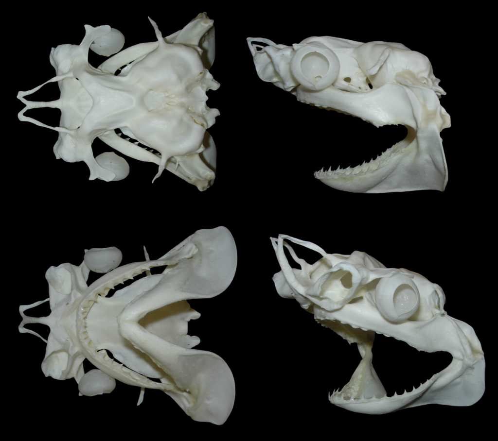
A couple of other projects we're currently working on is another full shark skeleton for a british aquaria, which should be completed very soon, and a few other critters as well, of which the most impressive is certainly this bowmouth guitarfish.

It's a small specimen that died in captivity. It's in good condition overall despite several issues with the skeleton, and shows some truly incredible morphological features.

Detail of the dentition. While many species have funky teeth, this species has always striked me as one of the most incredible chondrichthyan dentition.

Top view of the neurocranium during preparation. The cartilage was a bit damaged in the rostral area, but once again, really impressive thing. It's massive, and really looks like some bizarre hammerhead shark.

That's it for now, pictures of the finished specimens will follow soon!


Saturday, April 18, 2015

Blacktip update

Quick update on a couple of fishy bits. The blacktip shark skull is now finished, and waiting for the rest of the skeleton. As I'm currently quite busy with other stuff it might be a while before I get the time to finish it, but most of it is now completely clean, and just needs to be dried properly.

Carcharhinus melanopterus skull, finished.

Partial angelfish skeleton (probably Pomacanthus sp.). The pins are holding the fins in place while they dry.

Mako shark (Isurus oxyrinchus) drying. The basihyal is missing on the picture.

Couple of chondrichthyan parts: Raja asterias pelvic fins, Carcharhinus melanopterus hyoid arch, and Squalus acanthias skull and vertebrae.

Sunday, February 15, 2015

Fishy bits

Quick update on the blacktip (here)! I'm finally getting there. Still have to clean about half of the gills, but otherwise all pieces are cleaned, whitened, and most are dried already. I should have everything clean and dry by the end of the month so I can start putting the shark back together.

Dorsal and pectoral fins. As with the smooth-hound skeleton, I decided to keep the skin on most of the fins to protect the ceratotrichia.

Caudal fin, I used the same approach here.

And a couple of pictures from other projects:

Three catsharks (Scyliorhinus canicula) skulls in the cleaning process.

Degreasing a black grouper sull (Mycteroperca bonaci). This guy is incredibly greasy and will probably require multiple acetone soaks before whitening and final assembly.

Tuesday, December 23, 2014

WIP

Been a while since I posted anything, so here are a few pictures of various specimens in the works.

Recently I was lucky to get hold of a shortfin mako (Isurus oxyrinchus) head. It's a small individual, but I was very happy about it since I've been looking for this species for a long time. Since I got it during this year's comparative anatomy practicums, I also used the opportunity to bring it to the lab, which was a lot of fun. I then did the rough part of the cleaning process when the course was over.

Speaking about chondrichthyan jaw suspension.

The cleaned skull during the whitening process.

It's been a while since I macerated anything, but I kind of forgot several specimens in the maceration tanks which are now piling up and waiting to be degreased.

Various mammal and reptile skulls waiting to be degreased.

Still in the fish department, I was also able to get a couple of really cool-but tiny heads. Without the complete body it's hard to be sure but those are probably some kind of barbelled dragonfish (most probably in the genus Stomia). They are not in very good shape and have been kept in ethanol for a while but I'm currently in the process of cleaning the skull, more on that when I'm done with them.

Barbelled dragonfish head (Stomias sp.). Awesome little critter.

I've also been skinning and drying a couple of specimens for beetle work. We have some cool facilities at the lab to dry sediments that turned out to be very useful to dry critters as well.

Blue dot grouper (Cephalopolis taeniops). Found this one at the local fish store and ate it before saving the skeleton. Very tasty, and beautiful fish that should make a nice little skeleton.

Couple of captive bred herps, a python and chameleon. It was the first time I dissected one of these and they have a fantastic anatomy. Really looking forward to work with the skeleton of this one.

And finally, still playing around with clearing and staining specimens. Still have to get better at this, but I recently completed a batch of snakes so thought I would share a pic. Hopefully I will soon have other cool cleared specimens to show. And special thanks to a friend at the lab who gave me a hand skinning all these, and more :)

Cleared and stained cornsnakes (Pantherophis guttatus) and hognoses (Heterodon nasicus).


Friday, October 17, 2014

Blue-tongued skink skeleton

Some time ago I was asked by a french veterinarian to clean and articulate a complete blue-tongued skink skeleton (Tiliqua scincoides). The specimen had been autopsied, and the ribcage was damaged in the process. It was not in very good condition when I got it, especially the tail which was badly decomposed. For some reason the last caudal vertebrae had turned soft and spongy, and I could not save them, not sure what caused this. Here are a few pictures of the articulation process.

Sorting out the ribs


Almost done.


Finished skeleton

I usually reconstruct the sternal ribs using wire, but in this case I decided against it since most of the sternal region was missing in the first place (interclavicle, sternum, xiphisternum and sternal ribs were mostly destroyed during the autopsy). I did however reconstruct the missing suprascapulae and am rather pleased with how they turned out.

Here are a couple of pictures of the finished skeleton:

Dorsal view

The yellow spot on the hands and feet are remaining ligaments that didn't whiten very well.

Overall a very cool skeleton to work on, looking a bit like a reptilian version of the daschund.

Tuesday, July 1, 2014

Blacktip shark

I recently started to work on a very exiting project: a complete blacktip reef shark (Carcharhinus melanopterus), an aquaria casualty which was donated to the lab a few years ago and which has been sitting in the freezer since then. It's a small male (under 1 m long) and the head was cut off a few years ago as well, probably due to storage issues. Fortunately (and contrary to what is suggested by the picture below), the branchial apparatus was not-or only very little damaged in the process. However the scapulocoracoid is cut in half.

Head of the specimen as it was stored.

I took care of the head first. Cleaning was pretty straightforward using the method I already outlined on this blog (more detailed protocol to come later). I saved the scleral capsules, optic pedicels, and the complete branchial apparatus, including the extra branchials.

Cleaned and wet neurocranium. Note the optic pedicel (arrows) which articulates with the scleral capsules and help to move and stabilize it. Scale bar: 1cm.

The optic pedicels had to be removed since they tend to dry poorly. I treated them separately and will reattach them later.

Scleral capsule. Scale bar: 0,5cm. The hole is the optic foramen.

The scleral capsules are often completely overlooked on chondrichthyan skeletal preparations, which is a shame since they are very cool and interesting little pieces of cartilage.

Finished neurocranium. a: dorsal view, b: palatine view, c: lateral view. Scale bar: 1cm. The optic pedicels have been removed to be dried separately, due to their tendency to shrink and warp badly.

So far this specimen is a delight to work with since it is showed very little warping and shrinking (the main issues when dealing with cartilage skeletons) compared to other chondrichthyan species I've worked on (more on that later) during the drying process.

Neurocranium and jaws being coated with Paraloid B-72.

I usually don't coat my finished specimens, be they bone or cartilage, with anything since I am very careful with them, and most are stored either in specimen boxes or in display cases. However since this shark is going to be part of the lab teaching collection (we don't have a museum over here) I felt it needed to be properly protected. To do this I soaked or coated every piece of cartilage with Paraloid B-72, an acrylic resin soluble in acetone, which can be used either as an adhesive or as a consolidant on a wide variety of specimens, from skulls to ceramic objects.

 The rest of the body, still frozen.

About half of the gill apparatus is now clean and dry as well, so I just need to clean the rest and rearticulate everything. Next will be the rest of the body, which should be pretty straightforward, since it's basically only fins and vertebrae. More pictures later, so check back for updates on this specimen.

Saturday, May 10, 2014

Rattlesnake

I'm currently working on a large (c. 1.3 m) timber rattlesnake (Crotalus horridus) for a veterinarian. The specimen was in great shape, and I didn't notice anything special when I dissected and skinned it prior to cleaning. However, once cleaned I was very surprised by the bony growth present on numerous vertebrae, sometimes completely locking them together. I still have to do some research, but from what I read and discussed with a couple of people, they might be some kind of cancerous lesions. The skull also exhibits minor deformations.


Multiple views of various vertebral segments. Only the most serious deformities are pictured here. Scale bar=1 cm.


Several views of the articulated vertebral column.

The right dentary also exhibits slight deformation.


Thursday, April 10, 2014

Hedgehog take two

Remember the hedgehog skeleton I posted a few months ago? Well, it was badly damaged by careless students soon after. This was partly my fault because I articulated with as little wire as possible (all carefully hidden) and no additional support, for a free standing finished mount despite the knowledge that it would be used in zoology practicums and it wasn't meant to be a display only specimen.
That being said, it's also due to the incredible carelessness of some students when handling natural history specimens. The need to handle those properly and carefully, especially when it comes to teaching collections which are used by generation of students, cannot be stressed enough.
Well, lesson learned, and I spent some time repairing it and reinforcing it with wires and additional support. Unfortunately, a toe was lost in the process.

I also added a brass plaque to the base, which always adds to the finished piece I think.

 Right side view

 Detail of the scapular belt area

 Detail of the right leg


Despite the fact that I don't like at all to come back on a specimen I consider finished, this was an interesting experience, since I usually don't build skeletons with that much wiring although I kind of like the look of it.

It's now back in the university teaching collections, let's hope it stays in one piece this time!