I don't normally feature my own academic research on this blog, but since this is directly related to my interests in preparation, I thought I would mention the following paper:
A complete protocol for the preparation of chondrichthyan skeletal specimens. S. Enault, C. Auclair, S. Adnet and M. Debiais-Thibaud.
that was published a couple of months ago in the Journal of Applied Ichthyology. Of course, my own methods are more or less continually evolving as I like to refine my own protocols and experiment with new things, but given the scarcity of literature dealing with this subject, I think it might be of interest to some people.
Showing posts with label Fish. Show all posts
Wednesday, June 15, 2016
Saturday, June 4, 2016
Current projects
Still busy with our upcoming website, but in the meantime we have been working on a couple of really interesting projects, which are almost completed now, but I'll just post some early pictures for now.
We have been asked to clean a full shark and skate skeletons for a french natural history museum. This is a really exciting opportunity, and they look fantastic so far. The species are a school shark (Galeorhinus galeus) and undulate ray (Raja undulata). Both are male specimens caught off the coast of Brittany in France. The plan is to have them displayed next to their taxidermied versions, which were recently completed by Bernard Bourles, an excellent ichthyotaxidermist (his website can be found here: http://www.poissons.net/).
Galeorhinus galeus. This specimen is a male around 1.20 m long.
Dissection of the gills, this part needs to be done very carefully as not to damage the delicate branchial rays and extrabranchials, which are visible on top of the gills here.
Raja undulata, male specimen as well. This is the largest skate I've been working on so far.
As with most skate, many parts of the body are covered with various kind of spines and thorns. These are located on the pectoral fins and make handling such a specimen a bit tricky as they are extremely sharp.
The tail is also covered with them.
Massive jaw musculature.
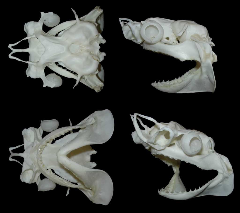
A couple of other projects we're currently working on is another full shark skeleton for a british aquaria, which should be completed very soon, and a few other critters as well, of which the most impressive is certainly this bowmouth guitarfish.
It's a small specimen that died in captivity. It's in good condition overall despite several issues with the skeleton, and shows some truly incredible morphological features.
Detail of the dentition. While many species have funky teeth, this species has always striked me as one of the most incredible chondrichthyan dentition.
Top view of the neurocranium during preparation. The cartilage was a bit damaged in the rostral area, but once again, really impressive thing. It's massive, and really looks like some bizarre hammerhead shark.
That's it for now, pictures of the finished specimens will follow soon!
Monday, May 25, 2015
Portuguese fish!
A few weeks ago I attended the 4th IAFSB (Interdisciplinary Approaches in Fish Skeletal Biology) meeting in Tavira, Portugal. I had a fantastic time there with great science and great people, and it came as a very nice surprise to see so many shark people there.
A few of us also took the opportunity to go to the local fish market see if we could find some cool fish to take home, which was really funny because for once I was not the only weirdo looking for heads or strange species. We saw loads of black and silver scabbardfishes and really wish I had gotten some of these, but I was already loaded with the cartilaginous stuff. Here's a few pictures of the specimens I brought back.
Torpedo sp. and Mustelus mustelus (? could be M. asterias, not quite sure on this one).
Couple of skates. The big one on the left is Raja microocelata and still have to ID the two smaller ones, probably Leucoraja something.
The torpedo skull on the left is very damaged: the fishermen cut through the jaws, one hyomandibular, and they also damaged the nose area (the antorbital cartilages are entirely missing). I still took it since it's the first time I come across torpedoes and it should still be useful as a practice specimen since I'm not very familiar with their skeletal anatomy.
Couple of skates. The big one on the left is Raja microocelata and still have to ID the two smaller ones, probably Leucoraja something.
Unidentified torpedo head (T. marmorata or T. torpedo) and whole T. torpedo.
The head is either from T. marmorata or T. torpedo. The first specimen had already been skinned and cut in pieces so ID is difficult, and they had both species at the market. The atlantic torpedo (Tetronarce nobiliana) is also present in the area, but this species is much bigger and usually found at greater depths. The complete specimen should make a very cool skeleton, unfortunately the pelvic fins are a little damaged.
Photo: Callie Crawford
Since none of us planned to be working on fresh specimens we had to improvise a bit. I had to buy several kilos of salt to prep them for the return trip. The other guys used some kind of cheap vodka and big plastic jars for their stuff. Wish I had a picture of this since it was quite funny. But the best part was trying to dissect fish using a table knife and nail clipper. Not the easiest way to do it!
Busted torpedo skull close up.
Photo: Callie Crawford
Everything is now safely stored in the freezer. I'll post WIP pictures here as I get around working on them.
Saturday, April 18, 2015
Blacktip update
Quick update on a couple of fishy bits. The blacktip shark skull is now finished, and waiting for the rest of the skeleton. As I'm currently quite busy with other stuff it might be a while before I get the time to finish it, but most of it is now completely clean, and just needs to be dried properly.
Carcharhinus melanopterus skull, finished.
Partial angelfish skeleton (probably Pomacanthus sp.). The pins are holding the fins in place while they dry.
Mako shark (Isurus oxyrinchus) drying. The basihyal is missing on the picture.
Couple of chondrichthyan parts: Raja asterias pelvic fins, Carcharhinus melanopterus hyoid arch, and Squalus acanthias skull and vertebrae.
Sunday, February 15, 2015
Fishy bits
Quick update on the blacktip (here)! I'm finally getting there. Still have to clean about half of the gills, but otherwise all pieces are cleaned, whitened, and most are dried already. I should have everything clean and dry by the end of the month so I can start putting the shark back together.
And a couple of pictures from other projects:
Dorsal and pectoral fins. As with the smooth-hound skeleton, I decided to keep the skin on most of the fins to protect the ceratotrichia.
Caudal fin, I used the same approach here.
And a couple of pictures from other projects:
Three catsharks (Scyliorhinus canicula) skulls in the cleaning process.
Degreasing a black grouper sull (Mycteroperca bonaci). This guy is incredibly greasy and will probably require multiple acetone soaks before whitening and final assembly.
Tuesday, December 23, 2014
WIP
Been a while since I posted anything, so here are a few pictures of various specimens in the works.
Recently I was lucky to get hold of a shortfin mako (Isurus oxyrinchus) head. It's a small individual, but I was very happy about it since I've been looking for this species for a long time. Since I got it during this year's comparative anatomy practicums, I also used the opportunity to bring it to the lab, which was a lot of fun. I then did the rough part of the cleaning process when the course was over.
Still in the fish department, I was also able to get a couple of really cool-but tiny heads. Without the complete body it's hard to be sure but those are probably some kind of barbelled dragonfish (most probably in the genus Stomia). They are not in very good shape and have been kept in ethanol for a while but I'm currently in the process of cleaning the skull, more on that when I'm done with them.
I've also been skinning and drying a couple of specimens for beetle work. We have some cool facilities at the lab to dry sediments that turned out to be very useful to dry critters as well.
And finally, still playing around with clearing and staining specimens. Still have to get better at this, but I recently completed a batch of snakes so thought I would share a pic. Hopefully I will soon have other cool cleared specimens to show. And special thanks to a friend at the lab who gave me a hand skinning all these, and more :)
Recently I was lucky to get hold of a shortfin mako (Isurus oxyrinchus) head. It's a small individual, but I was very happy about it since I've been looking for this species for a long time. Since I got it during this year's comparative anatomy practicums, I also used the opportunity to bring it to the lab, which was a lot of fun. I then did the rough part of the cleaning process when the course was over.
Speaking about chondrichthyan jaw suspension.
The cleaned skull during the whitening process.
It's been a while since I macerated anything, but I kind of forgot several specimens in the maceration tanks which are now piling up and waiting to be degreased.
Various mammal and reptile skulls waiting to be degreased.
Still in the fish department, I was also able to get a couple of really cool-but tiny heads. Without the complete body it's hard to be sure but those are probably some kind of barbelled dragonfish (most probably in the genus Stomia). They are not in very good shape and have been kept in ethanol for a while but I'm currently in the process of cleaning the skull, more on that when I'm done with them.
Barbelled dragonfish head (Stomias sp.). Awesome little critter.
I've also been skinning and drying a couple of specimens for beetle work. We have some cool facilities at the lab to dry sediments that turned out to be very useful to dry critters as well.
Blue dot grouper (Cephalopolis taeniops). Found this one at the local fish store and ate it before saving the skeleton. Very tasty, and beautiful fish that should make a nice little skeleton.
Couple of captive bred herps, a python and chameleon. It was the first time I dissected one of these and they have a fantastic anatomy. Really looking forward to work with the skeleton of this one.
And finally, still playing around with clearing and staining specimens. Still have to get better at this, but I recently completed a batch of snakes so thought I would share a pic. Hopefully I will soon have other cool cleared specimens to show. And special thanks to a friend at the lab who gave me a hand skinning all these, and more :)
Cleared and stained cornsnakes (Pantherophis guttatus) and hognoses (Heterodon nasicus).
Wednesday, October 29, 2014
Half digested baby barracuda skull
Well, maybe not quite exactly, but close. It's certainly partially digested, and I'm quite sure it's a sphyraenid, though it might be difficult to get a more precise ID based on its morphology. This guy was found in the stomach of a nicely sized small-spotted catshark (Scyliorhinus canicula) during last week comparative anatomy practicum. It's always interesting to see students' reactions when offered to investigate stomach contents, some are totally grossed out, but quite a few get really excited about it. We found all sort of things this week, mainly crustaceans, worms and small fish. I remember when I performed this same dissection as an undergrad a few years ago and found a large squid in the stomach of my specimen, a large female. I still have the beak I extracted from the half digested meal and the eggs I removed from the uterus laying around in a jar somewhere.
Partially digested sphyraenid head. Yum.
Thursday, October 23, 2014
Catshark breeding
For once this post will not feature too much dead stuff, but there will still be plenty of fish. We've had our own small catshark (Scyliorhinus canicula) breeding facility for a few months now. We currently have 15 mature individuals kept in a large tank at a nearby marine station, where they're happily reproducing and laying plenty of eggs.
The adults. They usually rest on the bottom of the tank but grow quite animated during feeding time.
Curious catsharks.
Plenty of eggs. Embryos are visible through many of them.
We bring the eggs to the lab every week or so, where we keep them in a large aquarium until the reach the desired developmental stage. The nice part is that although the eggcase color will vary from female to female, you can always see through it and check on the developing embryo inside.
Developing embryo.
We keep our weekly harvest in individual baskets in a large aquarium. The sausage shaped things on the bottom are tunicates (Ciona intestinalis). In the lower right corner you can see a newly hatched catshark resting on the bottom of the aquarium.
Catshark embryos at various developmental stages. From left to right: 1.5, 4 and 5 months old.
Scale bars: 0.4/0.6/0.8 cm
Saturday, October 11, 2014
Blue shark skull
This post is a bit overdue since this specimen has been completed for almost a year now, but I still wanted to write a few lines about it. It was my first large shark skull (and one of my first shark skulls for that matter, since I only started experimenting on numerous catshark specimens a few months later), and I made several mistakes in the process which I'll discuss a bit here.
I
found this specimen quite unexpectedly while out to buy some food
during the summer of 2013. I always check the fish department as well in
case they might have anything of interest, and got really excited when I
noticed the large shape on the stall. Although it's meat is sold on a
regular basis around here (I'm located on the Mediterranean coast) it
was the first time I saw the head in a shop, and the guys there were
nice enough to give me the head for free.
Blue shark (Prionace glauca) head.
Close up on the snout showing the ampullae of Lorenzini. The medial rostral cartilage is also visible through the skin.
Dissection of the head, with palatoquadrate and right optic capsule exposed.
The first mistake I made with this specimen was to attempt a complete dissection of the head. While it can be done, it often leads to broken structures in the skull, since in some areas the skin is a lot tougher than the cartilage underneath, especially around the rostrum and orbital processes. Doing so, I damaged one of the preorbital processes, which are fairly developed in this species.
The second mistake was to apply too much heat to the jaw which separated at the joints. Fortunately, the teeth didn't fall out, which is also a common occurrence when too much heat is applied. This is why you can't clean shark jaws by boiling them as Quinn is doing in Jaws.
Re-articulation of the jaw joints.
The jaws can be glued back in place, and on large skulls I use wire as well for additional strength. Even then, the jaws will be much more fragile and delicate than if the ligaments are preserved. The palatoquadrate and meckel's cartilage are carefully drilled and wired with brass wire and glued back together. The same process can be used for the hyomandibulars and to glue the jaw to the chondrocranium. On large specimens, I prefer to remove the jaw from the chondrocranium for proper cleaning. This way I can also apply heat to the chondrocranium without damaging the jaws.
Gluing the hyomandibulars back in place on a blacktip reef shark (Carcharhinus melanopterus).
Dorsal view of a blue shark skull, showing important distortion.
Anyway, it was still a very interesting specimen to work on. I hope to get my hands on a second one later one so I can clean it properly without repeating the same mistakes.
The finished specimen. I also kept the ceratohyals and basihyal and might glue them back later.
Wednesday, August 27, 2014
Greasy stuff
Grease is a prominent issue when
dealing with bones for a variety of reasons : conservation (the
grease might turn acidic, which can in turn damage the mineral part
of the bone structure. It can also provide a growth media for mold or
fungus.), aesthetic (greasy bones are usually yellow or brown,
unpleasant to look at and handle), smell (ever smelled a
cupboard/room full of greasy whales ?) and so forth. On the
other hand some people and museums go light on the degreasing due to
a belief that bones that have not been degreased hold together better
in the long run. While there might be some truth in this, I think
specimens must at least be superficially degreased to avoid to have
bones literaly oozing grease. The amount of grease present in a
specimen varies depending on a variety of factors: species, season,
age, sex, etc.
Here you can clearly see liquid grease oozing from the basioccipital area. This specimen was also beetled. Maceration usually gives a head start on degreasing over beetle cleaning but results in the disarticulation of the specimen.
I'm currently working on a couple of nicely greasy fish, so I
figured it would be a nice occasion to illustrate the issue.
Flathead catfish (Pylodictis olivaris)
First one is a partial flathead catfish skeleton, straight out of the beetles (not beetled by myself). It's not overly greasy, but you can still see the various shades of yellow and brown clearly asking for some degreasing.
American eel neurocranium (Anguilla rostrata), occipital view
Here you can clearly see liquid grease oozing from the basioccipital area. This specimen was also beetled. Maceration usually gives a head start on degreasing over beetle cleaning but results in the disarticulation of the specimen.
Mahi mahi (Coryphaena hippurus) neurocranium degreasing
I always use acetone to degrease fish material. It should be handled with care but it works very well for small vertebrates. Other degreasing techniques, especially a prolonged soak in warm water and detergent are more appropriate for larger specimens.
Friday, August 8, 2014
Pink teeth
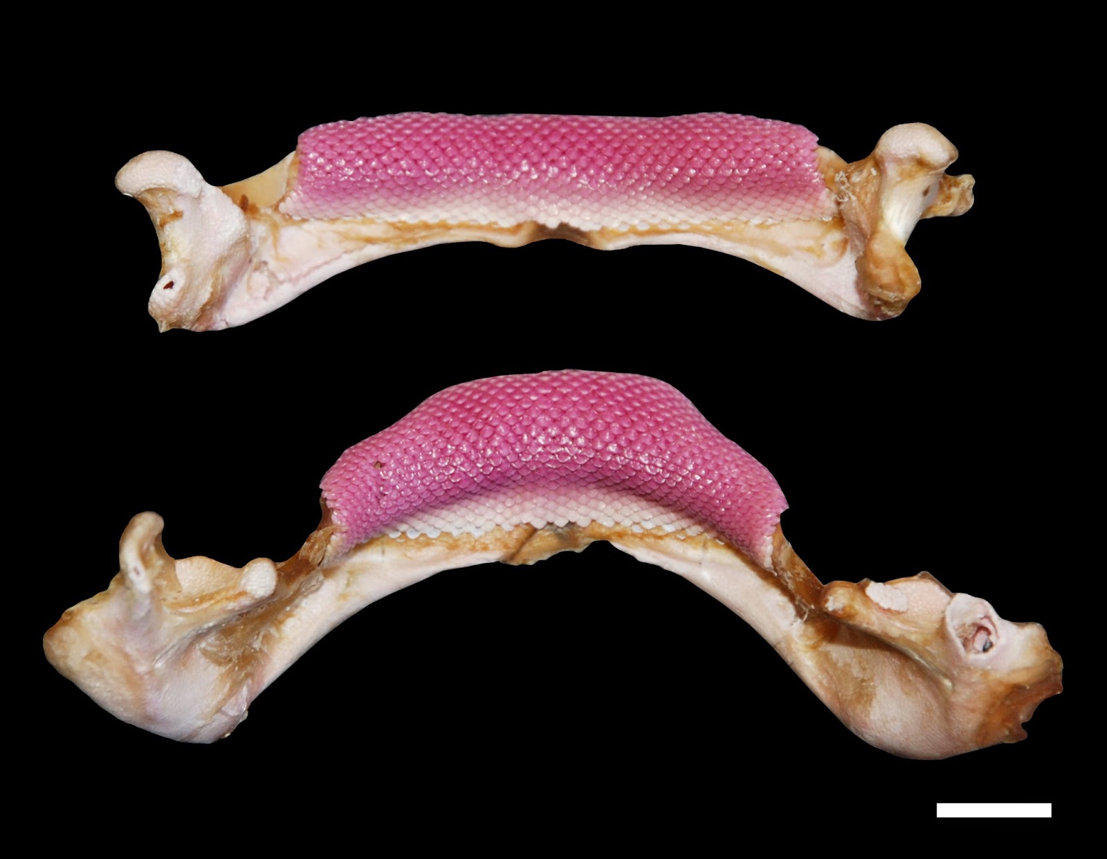
I recently came across this set of jaws from a Tortonese's stingray (Dasyatis tortonesei, which is probably invalid by the way) and was very surprised by the pink color of the teeth. It apparently not an uncommon occurrence in this species and is supposedly related to their diet, but I don't have any info/idea so far of what may be causing the the teeth to turn pink like that. For the little story, fishermen catching these apparently call them "whore rays" since they look to be wearing way too much lipstick.
And while doing my regular fish shop round the other day, I found a couple of these guys.
They are red bandfish, a small burrowing species found mainly in the eastern Atlantic and Mediterranean sea. First time I saw some despite them being regularly caught for those. They should have nice little skulls, and I might try to clear and stain a couple as well, more on that later.
D. tortonesei jaw, labial view. Scale bar: 1 cm
Red bandfish (Cepola macrophthalma) heads.
They are red bandfish, a small burrowing species found mainly in the eastern Atlantic and Mediterranean sea. First time I saw some despite them being regularly caught for those. They should have nice little skulls, and I might try to clear and stain a couple as well, more on that later.
Subscribe to:
Posts (Atom)


.jpg)
.jpg)